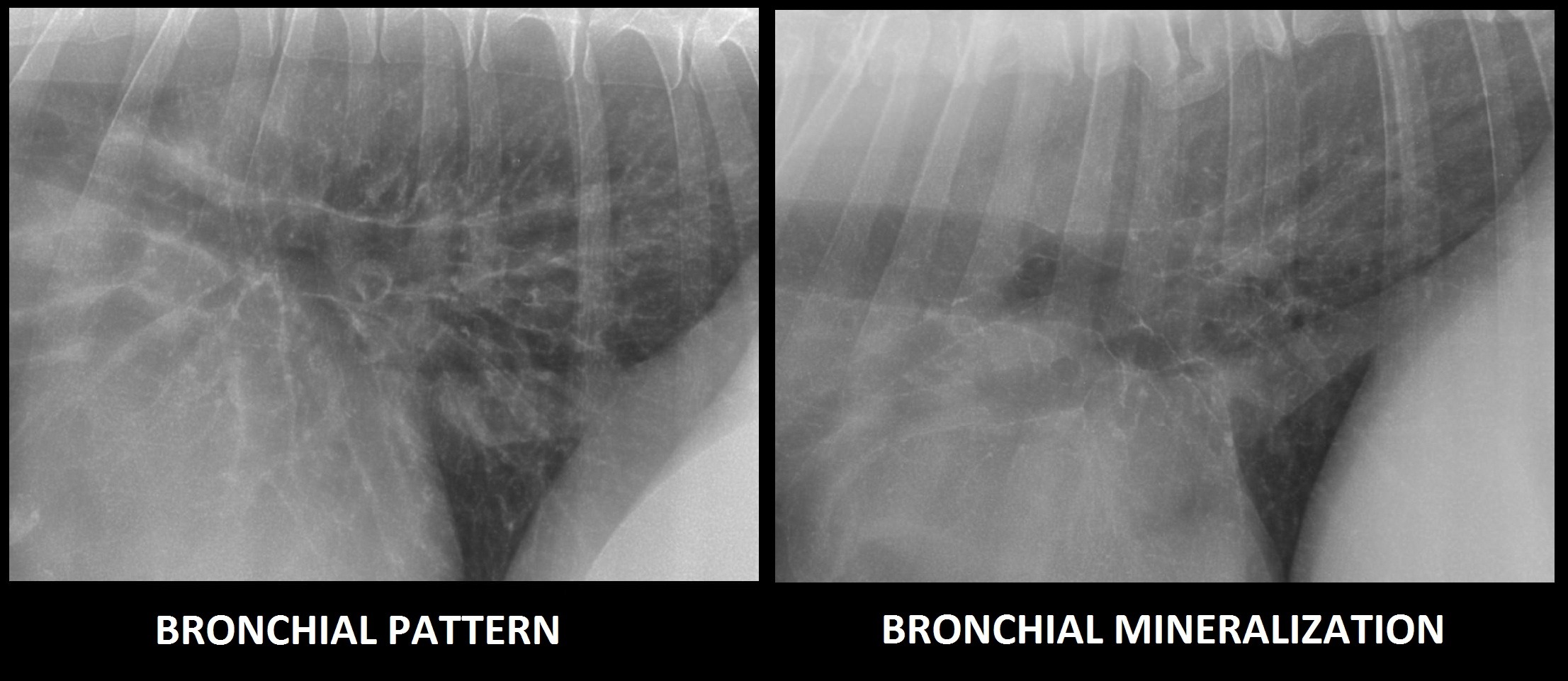
Alveolar Pattern Dog
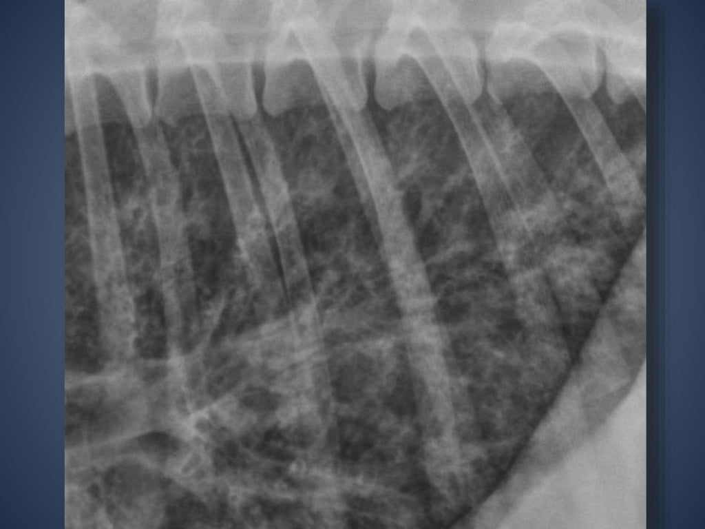
Alveolar Pattern Dog - Web left lateral thoracic radiograph of a dog with bronchopneumonia pneumonia. A particular form of the silhouette sign is the air bronchogram. Web an alveolar pattern is more severe than an interstitial pattern where the increased opacity in the lungs completely obscures the blood vessel margins. Alveolar lung pattern it is obtained when the air in the alveoli is substituted by material with higher density. Air bronchograms and lobar signs may also be present. An alveolar pattern is noted ventrally (right cranial and right middle lung lobes). It can be a subtle pattern to recognize, so lets look at some of the features. Web figure 1.photographs and diagnostic images (ct) revealing nature and extent of lesion. Web many patients may have a mixed pattern of breathing characterized by increased inspiratory and expiratory effort, as the disease processes may involve concurrent airway obstruction and altered lung compliance. Web an alveolar lung pattern is an opaque lung that completely obscures the margins of the pulmonary blood vessels. Underlying causes include viral infection, aspiration injury, foreign body inhalation, and defects in clearance of respiratory secretions. Web left lateral thoracic radiograph of a dog with bronchopneumonia pneumonia. Web the lung pattern you are dealing with is an alveolar lung pattern. Air bronchograms and lobar signs may also be present. Web thoracic radiographs revealed an alveolar pattern in the left cranial and caudal lung lobes, consistent with pneumonia. 3d reconstruction skull ct images show the nasomaxillary defect (yellow arrows) from the right lateral view (c), left lateral view (d), and dorsal view (e).also note the alveolar bone loss of left maxillary. Differential diagnoses for alveolar patterns are similar to those for interstitial patterns. This condition is caused by collapsed alveoli or infiltration (cellular or fluid types) of the alveolar lumen, which results in a consolidated increased opacity in the affected portion of the lungs. It can be a subtle pattern to recognize, so lets look at some of the features. An alveolar pulmonary pattern is created when the air within the alveoli is replaced with a material having a higher physical density, thus increasing the radiographic opacity of lung. A particular form of the silhouette sign is the air bronchogram. Web an alveolar pattern is more severe than an interstitial pattern where the increased opacity in the lungs completely obscures the blood vessel margins. Web many patients may have a mixed pattern of breathing characterized by increased inspiratory and expiratory effort, as the disease processes may involve concurrent airway. Following stabilization of the patient with oxygen, radiography plays a very valuable role in. Air bronchograms and lobar signs may also be present. Craniodorsal view (a) and left craniolateral view (b). Web many patients may have a mixed pattern of breathing characterized by increased inspiratory and expiratory effort, as the disease processes may involve concurrent airway obstruction and altered lung. Matthew winter, dacvr will review the radiographic features of lung patterns in dogs and cats as well as the keys to interpreting the meaning of these patterns. Upper and lower airway disease is common in dogs and cats, which can present with similar signs regardless of the location. This manifest as the inability to see margins of heart, vessels or. Lateral thoracic radiograph from a dog showing an unstructured interstitial pattern. Differential diagnoses for alveolar patterns are similar to those for interstitial patterns. This condition is caused by collapsed alveoli or infiltration (cellular or fluid types) of the alveolar lumen, which results in a consolidated increased opacity in the affected portion of the lungs. Web a bronchial and bronchointerstitial pattern. Web a bronchial pattern on radiographs indicates a condition that involves the airways. Uniform soft tissue opacity, the presence of air bronchograms, a lobar sign, border effacement with the heart or diaphragm and border effacement with the pulmonary vessels and outer serosal wall of. Web the alveolar pattern is indicative of lack of air in the alveoli. Furthermore, within the. The patient was hospitalized for supportive care and received iv fluids, cough suppressant, and antibiotic therapy (ie, enrofloxacin, doxycycline). Upper and lower airway disease is common in dogs and cats, which can present with similar signs regardless of the location. Air bronchograms and lobar signs may also be present. Web because the changes seen on thoracic radiographs are often indicative. Web the lung pattern you are dealing with is an alveolar lung pattern. Web thoracic radiographs revealed an alveolar pattern in the left cranial and caudal lung lobes, consistent with pneumonia. The airways are made out of cartilage which is radiolucent, but they have some surrounding soft tissue structures that can make them visible. Uniform soft tissue opacity, the presence. Web radiologic features consistent with cardiac enlargement were present in all dogs. Furthermore, within the caudodorsal lung field, a bronchointerstitial pattern predominates. Web an alveolar lung pattern is an opaque lung that completely obscures the margins of the pulmonary blood vessels. Web a bronchial and bronchointerstitial pattern are the most common radiographic lung patterns seen in canine eosinophilic bronchopneumopathy with. Air bronchograms and lobar signs may also be present. A total collapse of the alveoli (atelectasis) leads to a similar appearance. Web for the purpose of this article, we will focus on interstitial and alveolar patterns in our coughing and distressed patients, and touch on bronchial patterns. 3d reconstruction skull ct images show the nasomaxillary defect (yellow arrows) from the. Web radiologic features consistent with cardiac enlargement were present in all dogs. It can be a subtle pattern to recognize, so lets look at some of the features. Air bronchograms are visible extending into the right middle lobe. 3d reconstruction skull ct images show the nasomaxillary defect (yellow arrows) from the right lateral view (c), left lateral view (d), and. Web the components of an alveolar pattern include: Web a bronchial pattern on radiographs indicates a condition that involves the airways. The only distinction these patterns make with regards to clinically relevant information is the severity of the disease. Web left lateral thoracic radiograph of a dog with bronchopneumonia pneumonia. Matthew winter, dacvr will review the radiographic features of lung patterns in dogs and cats as well as the keys to interpreting the meaning of these patterns. Web figure 1.photographs and diagnostic images (ct) revealing nature and extent of lesion. Web the alveolar pattern is indicative of lack of air in the alveoli. Uniform soft tissue opacity, the presence of air bronchograms, a lobar sign, border effacement with the heart or diaphragm and border effacement with the pulmonary vessels and outer serosal wall of. Following stabilization of the patient with oxygen, radiography plays a very valuable role in. It can be a subtle pattern to recognize, so lets look at some of the features. Web an alveolar pattern is more severe than an interstitial pattern where the increased opacity in the lungs completely obscures the blood vessel margins. The airways are made out of cartilage which is radiolucent, but they have some surrounding soft tissue structures that can make them visible. An alveolar pattern is noted ventrally (right cranial and right middle lung lobes). Web for the purpose of this article, we will focus on interstitial and alveolar patterns in our coughing and distressed patients, and touch on bronchial patterns. Furthermore, within the caudodorsal lung field, a bronchointerstitial pattern predominates. Web typical differentials for interstitial and alveolar patterns in dogs include:
Thoracic radiography of a dog with pneumonic plague (case 2). Left

Radiographic Approach to the Coughing Pet • MSPCAAngell

Figure 6 from Distribution of alveolarinterstitial syndrome in dogs
Radiographic Approach to the Coughing Pet • MSPCAAngell
The Radiographic Approach to the Coughing Dog
Alveolar pattern or normal anatomy in the thorax of a young dog?

Imaging the Coughing Dog

Visual assessment of the classification results of a

Radiographic Approach to the Coughing Pet • MSPCAAngell

Imaging the Coughing Dog
Web Alveolar Pulmonary Pattern An Alveolar Pattern Is The Result Of Fluid (Pus, Edema, Blood), Or Less Commonly Cells Within The Alveolar Space.
Characterized By The Lobar Sign, Air Bronchograms And Border Effacement.
This Manifest As The Inability To See Margins Of Heart, Vessels Or Diaphragm.
Lateral Thoracic Radiograph From A Dog Showing An Unstructured Interstitial Pattern.
Related Post: